In modern time, several invasive surgeries that involve stereo-tactic placement of medical equipment, such as catheters, are being done blindly. This puts patients at a risk of the surgeon missing during incision of the catheter, like in some applications in neurosurgery. Modern research technologies have introduced the concept of hybrid reality to the field of medicine by superimposing medical images onto the patient to noninvasively visualize the internal anatomy of the body. It is believed that this may lead to remarkable advances in the field of medicine by providing the surgeons with more insight to strategically plan the invasive operation before surgery and also provide guidance for the surgeon during surgery.
The concept of hybrid reality
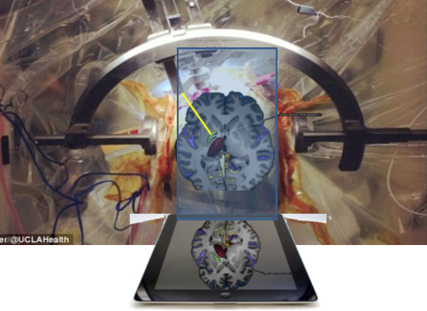
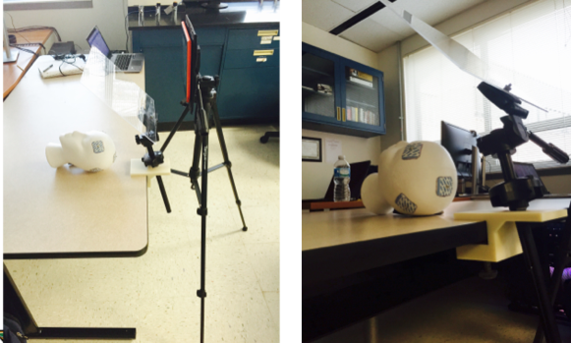
Dr. Mahesh Shenai, a neurosurgeon at the the INOVA hospital in Fairfax, VA, proposed to apply the concept of hybrid/augmented reality to neurosurgery. Ventriculostomy, a surgery done to remove excess cerebrospinal fluid buildup in the ventricles of the brain to maintain it’s intracranial pressure, is done by placing landmarks on the top of a patient’s head and assuming proper placement. This project proposes to build an optical device to superimpose images of a CT scan done on a cadaver head and develop software that displays and manipulates these images. The desired image chosen by Dr. Shenai, the neurosurgeon, is then projected onto the cadaver head. He will then attempt ventriculostomy on the cadaver head based on the developed system to measure percent accuracy of catheter incision
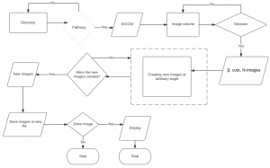
The flow of the developed software:
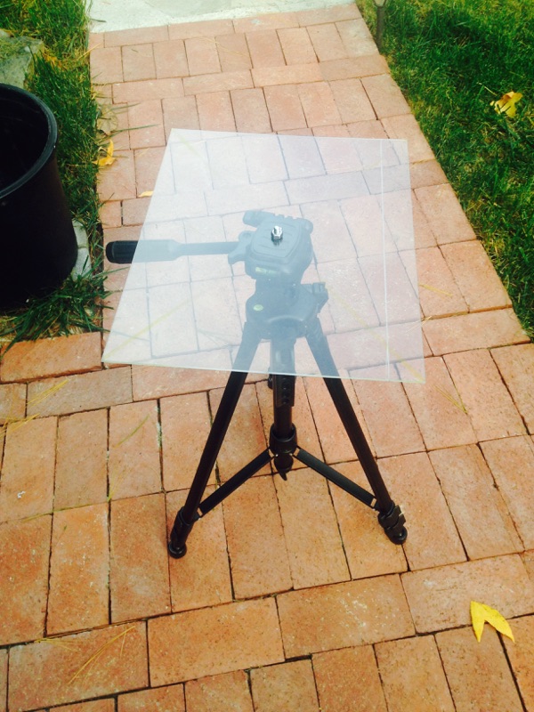
Initial design (first semester) for the optical device:
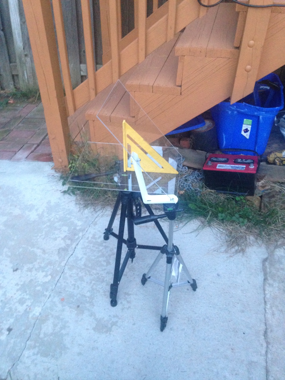
Final design (first semester) of the optical device:
Demonstration